facs flow cytometry protocol
Ive been using 1xDPBS to resuspend my cells for FACS analysis but lately Ive been getting bad results and my boss suggested that I used 1BSA. Bench Tools.

Flow Cytometry Sample Preparation Proteintech Group
Asked 16th Apr 2014.

. Add 100 μl of Fc block to each sample Fc block diluted in FACS buffer at 150 ratio. FSC-H for getting singlets from a. FACS is an abbreviation for fluorescence-activated cell sorting which is a flow cytometry technique that further adds a degree of.
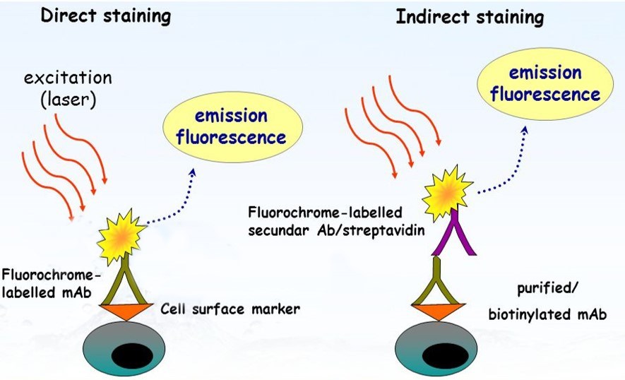
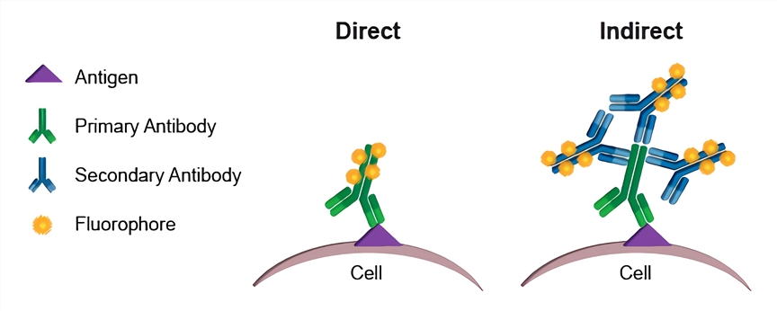
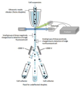
Propidium iodide is a suspected carcinogen and should be handled with care. Indirect labeling requires two incubation steps firstly with a primary antibody then with a compatible secondary antibody. Fluorescence-activated cell sorting is a specialized type of flow cytometry.
Please read the following cell viability protocol in its entirety before beginning. Biomarker Research via EXCEL. Add 01-10 μgml of the primary labeled antibody.
The dye must be disposed of. Incubate for at least 30 min at room temperature or 4C in. BD FACS Sample Prep Assistant SPA III.
I dont understand the concept behind FSC-A vs. It provides a method for sorting a heterogeneous mixture of biological cells into two or more containers one cell at. The samples should be resuspended in Cell Staining Buffer.
Moreover TagRFP657 was shown to be an efficient protein tag for the superresolution fluorescence imaging using a stimulated emission depletion microscope as well as for multicolor wide. Quality Assurance and Immunophenotyping of Lymphocytes. Cells are usually stained in polystyrene round bottom 12 x 75 mm 2 Falcon tubes.
There are several different dyes that can be used in these assays including propidium iodide PI 3 4 7-amino actinomycin-D 7-AAD Hoechst 33342 and 33258 and 46-diamidino-2-phenylindole DAPIFor example Chopra S et al labeled mouse bone marrowderived dendritic cells and paw single cell suspensions with 05 μgml DAPI from. This six-channel five colors and one FRET channel real-time PCR instrument combines advanced optical technology with precise temperature control to deliver sensitive reliable detection for singlexplex or multiplex reactions. Perform fluorescence activated cell sorting FACS or flow cytometric analysis.
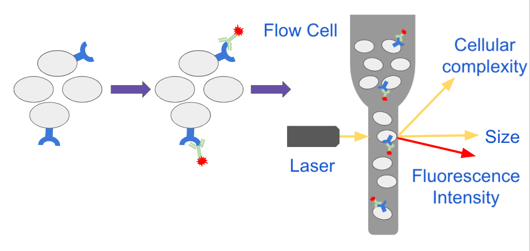
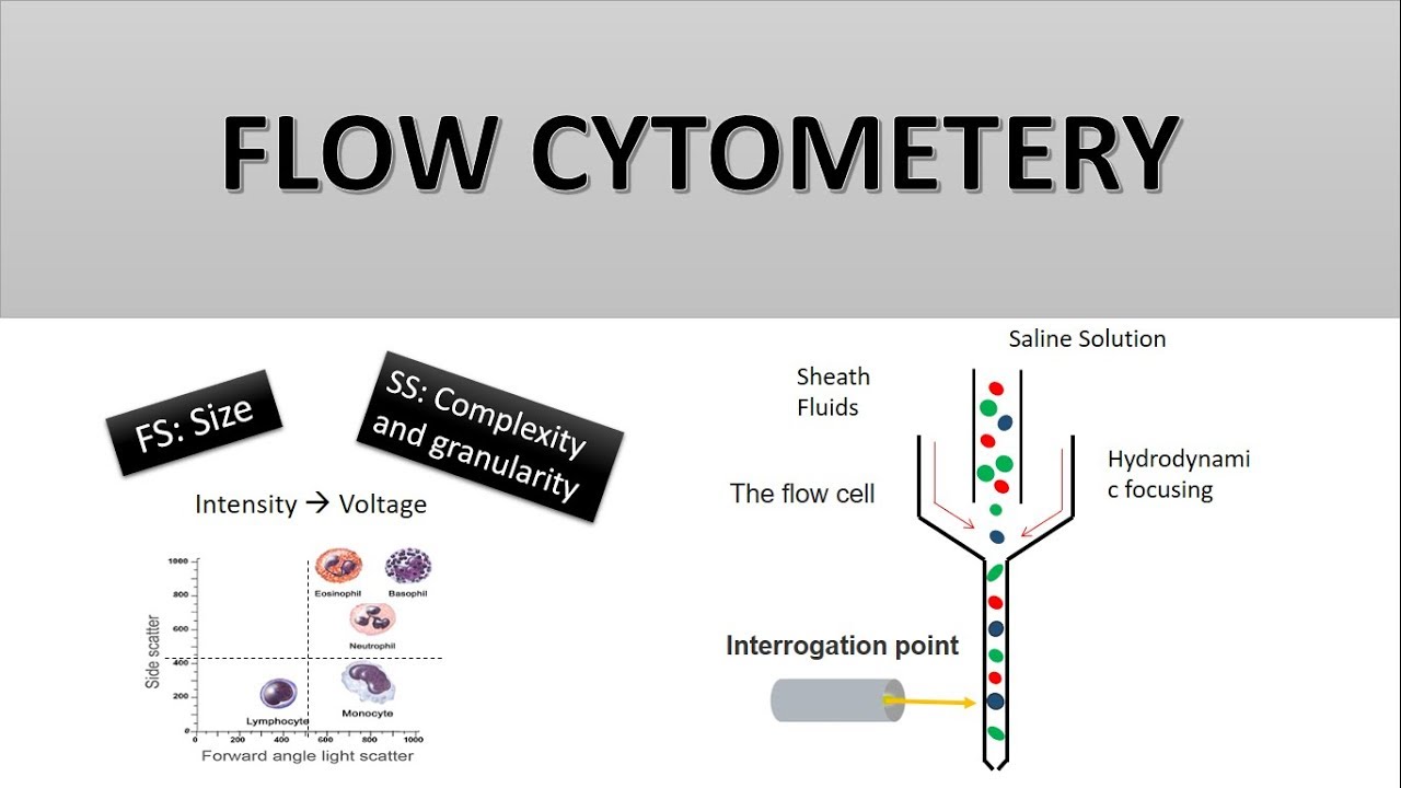
It is a technique that is used to detect specific proteins in the given sample. Download our membrane staining summary. The properties measured include a particles relative size relative granularity or internal complexity and relative fluorescence intensity.
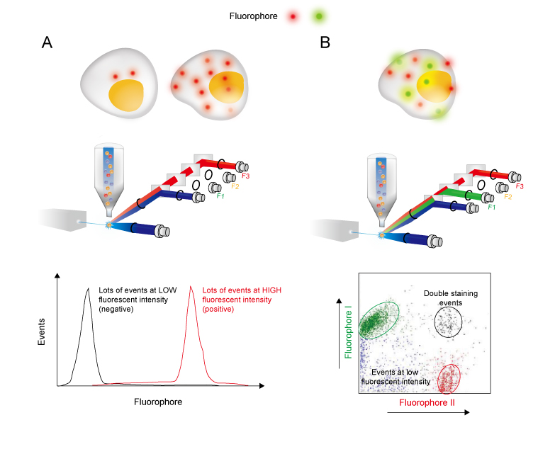
Harvest wash the cells and adjust cell suspension to a concentration of 1-5 x 10 6 cellsmL in ice-cold PBS 10 FCS 1 sodium azide. Flow cytometry multicolor experiments may need compensation when there is fluorescence spillover Figure 1Pairing fluorochromes based on antigen density fluorochrome brightness and separating by channels helps to minimize the effects from spillover and may remove the need for compensation from smaller experiments. Centrifuge at 1500 rpm for 5 min at 4C.
Flow cytometry has been extensively exploited in immunology hematology and oncology to define cell populations via intrinsic scatter properties cell surface antigen expression and other fluorescence parameters 1-3Our insights into blood lineage development and disease are a result to a significant degree of the continuous refinement of. There is no need to use sodium azide in these buffers it will. Flow Cytometry Panel Design Support Work with one of our technical sales specialists to discuss your experimental needs and guide you through the process.
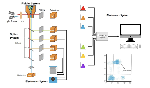
The CFX96 Touch System is a powerful precise and flexible real-time PCR detection system. However as the number of parameters and colors. Flow cytometry is a technology that simultaneously measures and then analyzes multiple physical characteristics of single particles usually cells as they flow in a fluid stream through a beam of light.
What cells are sorted by CD11bGR1 and CD11bGR1- flow cytometry. Flow Cytometry Support CenterFind technical support recommendations for your flow cytometry workflows including tips for experimental setup and in-depth troubleshooting help. BSA and FBS or any other serum for that matter will accomplish pretty much the same thing when staining cells for flow cytometry.
Dilutions if necessary should be made in FACS buffer. Incubate on ice for 20 min. However they can be stained in any container for which you have an.
Flow Cytometry is a technique used to detect and measure the physical chemical characteristics of a population of cells or particles Learn more. Print this indirect flow cytometry protocol. General procedure for flow cytometry using a primary antibody and conjugated secondary antibody.
It usually involves two major processes namely SDS-polyacrylamide gel electrophoresis and protein blotting and testing. General procedure for flow cytometry using a conjugated primary antibody. The Savvy Scientists Buffers Guide.
Show More Show Less. Expert Tips on IHC Optimization. Learn the concept behind Western blotting.
The following protocol has been developed and optimized by RD Systems Flow Cytometry Laboratory for cell viability staining using propidium iodide. Clinical Applications of Flow Cytometry. If you are unable to immediately read your samples on a cytometer keep them shielded from light and in a refrigerator set at 4-8C.
From flow cytometers and sorters for simple to complex research applications to an extensive selection of reagents tools educational resources and protocols we support you in navigating your multicolor flow cytometry workflow journey. The red-shifted absorbance allows for the excitation of TagRFP657 by the standard 633-640 nm red lasers used in flow cytometry analyzers and FACS instruments. And profile cells in a heterogeneous fluid mixture.

Macsquant Tyto Cell Sorting Applications Cell Sorter Macs Flow Cytometry Products Miltenyi Biotec Usa

Optimized Flow Cytometric Protocol For The Detection Of Functional Subsets Of Low Frequency Antigen Specific Cd4 And Cd8 T Cells Sciencedirect
Flow Cytometry Protocols

2015 We Have Developed A Simple Cost Effective And Labor Efficient Two Step Protocol For Preparing Adherent Cells For High Throughp Flow Cytometry Cell Flow

Flow Cytometry Creative Biolabs
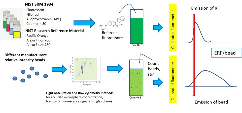
Quantitative Flow Cytometry Measurements Nist
Flow Cytometry Methodology
Direct Staining Flow Cytometry Creative Biolabs

Flow Cytometry Based Protocols For Human Blood Marrow Immunophenotyping With Minimal Sample Perturbation Star Protocols

Fundamentals Of Flow Cytometry Aat Bioquest

Flow Cytometry Facs Protocols Sino Biological
Analyzing Single Cells With Flow Cytometry
Flow Cytometry Creative Biolabs

Flow Cytometry Illustrated Assay Novus Biologicals

Flow Cytometry For Dna Analysis Youtube

In The Protocol Developed By Bernhard Fuchs S Team Bacterial Groups Are Enriched In Three Steps 1 In Situ Hybridization Postdoctoral Researcher Microbiology
How Does Flow Cytometry Work Nanocellect
The Principle Of Flow Cytometry And Facs 1 Flow Cytometry Youtube
Flow Cytometry Guide Creative Diagnostics